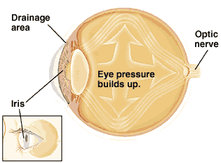


Let’s begin by drawing a fitting comparison between the human eye and a balloon so that it could be easier for you to understand the subject of “eye pressure”.
Visualize a balloon. See it filled with water. It goes without saying that the inward pressure exerted on the balloon will be determined by the volume of water that is pumped into it. This, in a nutshell, is what eye pressure is all about – the tissue pressure inside the eye which is determined by the balance between the production and drainage of the fluid inside the eye called the aqueous humor.
Intraocular pressure is the technical term that is used to refer to the measure of pressure in eyes, and the unit of measure is millimeters of Mercury (denoted as mmHg). A normal eye should have somewhere between 10-21 mm Hg; anything below or above that range should immediately raise a red flag.
There are a number of reasons why pressure behind eyes builds up. Here are some of them:
 Excessive aqueous production-The clear liquid that is produced in the eye is referred to as the aqueous humor. This liquid, produced by the ciliary body which is behind the iris, fills the anterior chamber of the eye (the space between the iris and cornea) and drains out of this chamber via trabecular meshwork around the periphery of anterior chamber. If too much of the aqueous humor is produced, then eye pressure increases.
Inadequate aqueous drainage-The opposite of excessive aqueous production is inadequate drainage of the aqueous humor. Slow drainage results in an imbalance of what is the ideal production and drainage quota of the aqueous humor. This results in the buildup of pressure in eyes because expulsion of the fluid is strained.
Trauma-Trauma to eyes can cause the buildup of pressure in eyes. Trauma, in this case, refers to any injurious impact on the eye. Trauma, as a cause of ocular hypertension, is tricky to identify because sometimes it might manifest itself after months or even years after impact occurred.
Medication-Certain medications can cause ocular hypertension, the most common being steroid medicines used to treat asthma and those used as eye drops.
Other possible causes of the increase in ocular hypertension are underlying eye conditions such as pigment dispersal syndrome or pseudo exfoliation syndrome, age (those who are above 60 are highly predisposed to ocular hypertension), and lastly a family history of this condition.
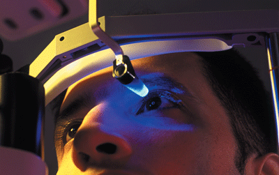
You may ask “how do I detect changes in eye pressure?” The fact is, you cannot detect pressure changes on your own. You will need to visit an eye specialist (ophthalmologist) who will administer tonometry tests. Below are the two types of tonometry tests available.
 Applanation tonometry-In this test, a tonometer is briefly placed on the cornea of the eye to flatten it. This test aims to analyze the amount of force needed to flatten the eye. To numb the eye, anesthetic drops are used so that the subject feels no pain.
Noncontact tonometry-This test basically estimates eye pressure, using a puff of air that blasts through the surface of the eye. It’s quick and there is no contact between any instrument and the eye.
After these tests are administered, one will only be subjected to more tests if one has abnormal intraocular pressure. Further tests are administered to check the risk of developing eye diseases.
There are three ways of treating ocular hypertension. They are as follows:
Medication-The medication used to deal with ocular hypertension is normally administered as eye drop. There are many prescriptions in the market and each will have a different dosage as well as possible side effects. One would do well to have a candid talk with their doctor before embarking on this journey.
Laser and Surgical Therapy-These two are not the first priority for many doctors because of the risks that are associated with both of them. It is important to note that laser treatment is used in the event that the patient cannot tolerate their medication.